16 de abril de 2024

Gyromitra sect. Discina in Europe


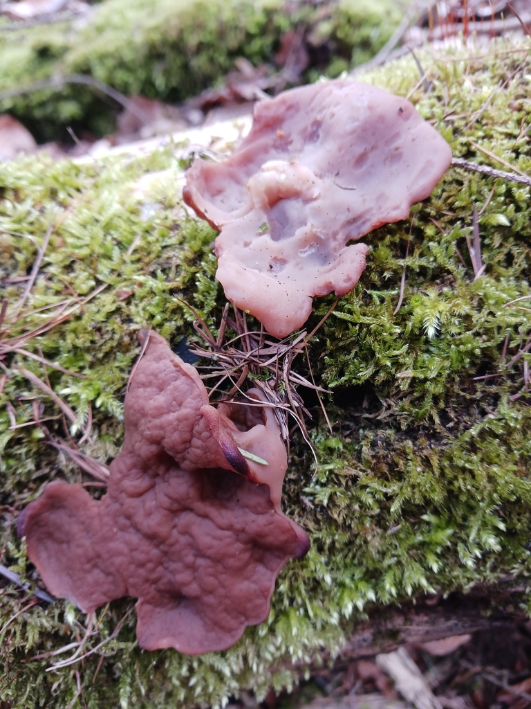
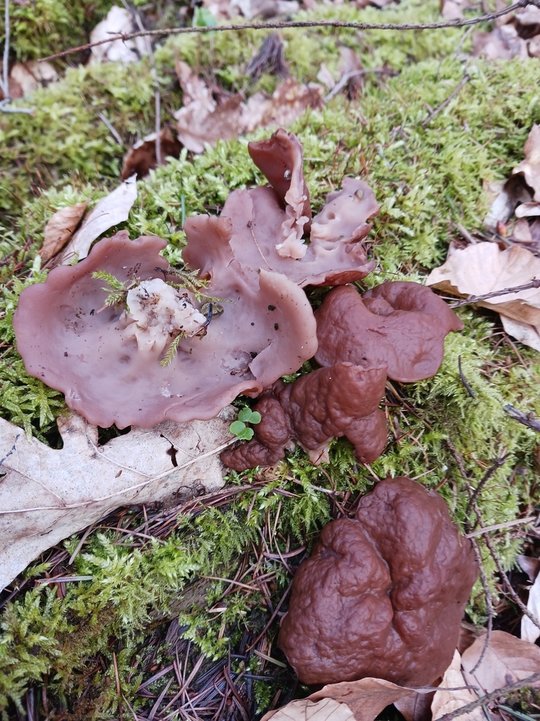
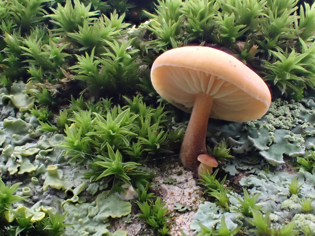
During spring, you might find so called "Pig's Ears" growing on dead wood or directly on the ground, brownish cup fungi that get spread out flat when ripe and belong to genus Gyromitra, the "False Morels". The problem with IDing Pig's Ears is that there are more than just one species and that those species look nearly identical to each other. In the past, it was quite easy to identify them to what was believed to be species level at the time: Pig's Ears growing on dead deciduous wood were called Gyromitra parma and those growing on dead conifers were called Gyromitra perlata (or Gyromitra ancilis; those two names are synonymous). But in 2009 it was shown by VAN VOOREN & MOREAU that the situation is unfortunately not that easy. Now there are about ten possible species in Europe: G. perlata (syn. G. ancilis), which is probably still the most common species, G. geogenia, G. intermedia, G. warnei, G. fluctuans, G. megalospora, G. olympiana, G. accumbens, G. leucoxantha and the mainly American species G. convoluta. Without microscopy, it is only possible to ID them to subgenus level on inaturalist: subgenus Discina. (In subgenus Discina there are two sections, sect. Discina and sect. Pseudogyromitrae with Pig's Ears belonging exclusively to sect. Discina, so you could ID them to section level, but as those sections do not currently exist on inaturalist, this information might be a bit superfluous. The information that subgenus Discina will sometimes be treated as its own genus, which makes the Pig's Ears Discina sect. Discina, might also be a bit superfluous, as inaturalist still considers Discina a subgenus of genus Gyromitra.)

If you have a microscope and want to ID them to species level, you can use the literature linked below. Unfortunately, I have not found an English key until now, only a typification of the most common species G. perlata (syn. G. ancilis). If you know an English key or any other useful current literature in your European language of choice, especially about distribution, please feel free so to post a link in the comments so I can add it to the literature list. Linked to this journal entry, there is also an observation of G. perlata including microscopic images you can compare with.

If you have valuable information to add, please do so in the comment section. Just because I'm writing this journal entry doesn't mean I'm the leading expert on those fungi.


Literature:

English:

French:

German:


Posted on 16 de abril de 2024, 03:45 AM by mangoblatt mangoblatt | 1 observação | 1 comentário | Deixar um comentário

22 de fevereiro de 2024

Sarcoscypha spp. in Europe


In late winter and early spring, beautiful red cup fungi of genus Sarcoscypha can be found growing on fallen twigs and branches. In Europe, there are three possible species (four if you include S. macaronesica, a species endemic to Macaronesia that was also found on the Balearic islands): S. coccinea, S. austriaca and S. jurana. Orange, yellow and white forms are also known from these species. Even though they all prefer slightly different substrates, there is some overlap. For instance, even though S. jurana seems to only grow on Tilia spp., S. austriaca, possibly the most abundant species in Temperate Europe (in the Mediterranean area, the most abundant species certainly is S. coccinea), has been reported to sometimes also grow on Tilia spp. It doesn't help that it is seldom possible to confidently identify the species of a fallen twig. (Some substrates seem to be unambiguous, though. S. austriaca is for instance the only species reported growing on Alnus spp., S. coccinea the only one on Quercus ilex and Pseudotsuga menziesii.) And macroscopic differentiation, well... The outside of S. coccinea tends(!) to be a bit less pale than the outside of the other two species, but not always, and it also may(!) have a slightly more hairy/spikey cap margin, but this might turn out to be a false generalisation, and the two other species do not differ macroscopically in any significant way from each other. Which means that unfortunately, you really cannot identify any one of the three species without microscopy.

But under the microscope, they are quite easily distinguished, at least while they are still fresh (if not, you may have to look for germinating spores, which can take for ages). Normally, you will just need to look for the distribution, amount and size of the oil droplets in fresh spores and if the small hairs on the outer surface of the cup are straight or curled up like a corkscrew. In the observations linked to this journal entry, you will find pictures of all relevant microscopic features of freshly collected specimens. In the literature section, you will find microscopic keys and further descriptions.

Exceptions: According to @epopov, the Saint Petersburg and Moskow area are devoid of S. coccinea and S. jurana: "In this particular area, my attempts to find Sarcoscypha coccinea or S. jurana for more than 20 years have not been successful. Among several hundreds herbarium specimens i've seen all reliable collections of other [non-austriaca] species of this genus in European part of Russia were made south of the latitude of Moscow, in the zone of broadleaved forests and further south in the Caucasus." (source) The same can be said for Estonia, as this study has found out, and for Finland, where according to @bodhiheera (see comments) a very active mycological community has looked "for ages" for S. coccinea, but found nothing. In my subjective(!) opinion that is sufficient evidence to rule out other Sarcoscypha spp. in the region between Estonia, Moscow and Finland, until verified findings of S. coccinea or S. jurana in the region appear (so I would still encourage microscopy). In my subjective opinion, every other place in Europe (with the exception of the Macaronesian islands with their endemic S. macaronesica, of course) still needs microscopic analysis.

If you have valuable information to add, please do so in the comment section. Just because I'm writing this journal entry doesn't mean I'm the leading expert on those fungi.


Literature:

Czech:

Dutch:

English:

French:

German:

Italian:

Polnish:

Spanish:

Swedish:

Ukrainian:

Posted on 22 de fevereiro de 2024, 09:36 PM by mangoblatt mangoblatt | 3 observações | 14 comentários | Deixar um comentário

24 de janeiro de 2024

Tremella mesenterica vs Naematelia aurantia


Tremella mesenterica surrounded by its common host Peniophora quercina

Mostly during winter, gelatinous fungi of yellowish to slightly orange colour can easily be found growing out of dead (or dying) wood. Even though they all look morphologically more or less the same, there are two distinct species in Europe: Tremella mesenterica and Naematelia aurantia.

Fortunately, no microscopy is needed to identify them, but you will have to look carefully at the dead wood they are growing on and check if you find their host organism. Because yes, even though those fungi grow on dead wood, they do not consume the wood, they consume another fungus. And that fungus can either have corticoid fruiting bodies, often greyish-brown, pale or purple in colour, and belong to genus Peniophora, or it can have effuso-reflexed fruiting bodies, often yellowish in colour, and belong to genus Stereum (as far as I know, it is always Stereum hirsutum).

In short (you'll find an observation attached to this journal entry for every one of those three cases):

  • If the fungus is found next to Peniophora sp, it can be identified as Tremella mesenterica.
  • If the fungus is found next to Stereum sp, it can be identified as Naematelia aurantia.
  • If neither or both are found on the same piece of wood, the fungus can only be identified as belonging to order Tremellales.

Even though many people ID those fungi as T. mesenterica if Stereum sp. is not visible on the photograph, you really should NOT do this... with one possible excpetion: On very thin, long twigs not or only very distantly connected to thicker branches, T. mesenterica can be inferred, because those are a good habitat for Peniophora sp. but not for Stereum sp. In this case you may ID to species lvl without a visible host. Generally, T. mesenterica will look a bit more glossy and transparent than the rather dull N. aurantia, but this should only be used as a supportive criterion to your ID, especially because, at least in my opinion, it is sometimes difficult to tell if something is glossy or humid or if it's just the lighting.

So please, if you want your observation of a yellowish, gelatinous fungi growing on dead (or dying) wood to count, provide a picture of that fungi together with its host. Look for it on the piece of wood you're photographing, especially on the side opposing the fungus. More often than not you will be able to spot the host after a short search. Please also take a look at the observation of N. aurantia linked below. You'll see that it would've been really easy to overlook the host, if I hadn't looked for it carefully, and you'll also see that if I hadn't, following the rule of thumb "No Stereum visible: T. mesenterica it is" would have let to a misidentification of the specimen. In a comment to this journal entry you will find some more N. aurantia observations that could have been (or have been) esily misidentified as T. mesenterica.

If you have valuable information to add, please do so in the comment section. Just because I'm writing this journal entry doesn't mean I'm the leading expert on those fungi.


Primary literature:

Posted on 24 de janeiro de 2024, 05:33 PM by mangoblatt mangoblatt | 3 observações | 3 comentários | Deixar um comentário

10 de janeiro de 2024

Flammulina spp. in Europe


In Europe, there are 8 (9 if you count Laccariopsis mediterranea, formerly F. mediterranea) well established Flammulina species: F. elastica, F. velutipes, F. populicola, F. rossica, F. fennae, F. finlandica, F. ononides and F. cephalariae. (It may also be possible, though very unlikely, that F. filiformis may have escaped cultivation in some places and is found wild locally.) With exception of the host specific and very rare F. ononides on Ononis spinosa, F. cephalariae on Cephalaria leucantha and Laccariopsis mediterranea on Ammophila litoralis, microscopy is always needed to differentiate between those species. Especially important are spore sizes and Q (ratio of spore length to width). In most cases, the pileipellis should be observed, too.

If you have valuable information to add, please do so in the comment section. Just because I'm writing this journal entry doesn't mean I'm the leading expert on those fungi.


Primary literature:


Secondary literature:

Dutch:

English:

Finnish:

French:

German:

Italian:

Posted on 10 de janeiro de 2024, 07:37 AM by mangoblatt mangoblatt | 2 observações | 3 comentários | Deixar um comentário

Arquivos